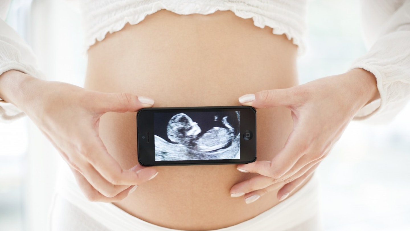
Each pregnancy ultrasound scan is pretty exciting (you get to see your baby) and slightly scary (just what will you see?), so it’s a good job to get prepared.
Chances are you’ve squinted at a friend’s grainy ultrasound picture, nodded and wondered just exactly what you were looking at. Now it’s your turn for the scans and excitement.
Here's a guide to all the routine scans during pregnancy...
How do ultrasounds work?
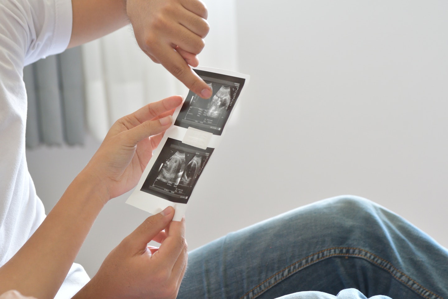
You get what ultrasounds do, but how do they work? Impress your partner by understanding the lingo.
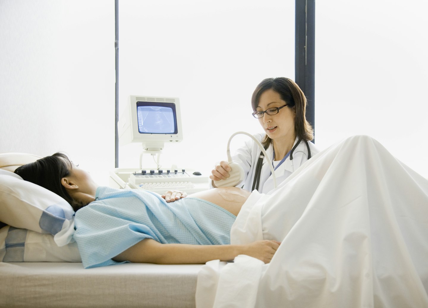
Firstly, the device the sonographer holds against your bump is called a transducer.
This sends high-frequency sound waves in your abdomen, where your baby’s tucked away.
These waves bounce off your baby and back to the computer to be translated into a picture, that’s what comes out as the white area.
"You could be given extra scans in your pregnancy if particular issues are a concern," says Dean Meredith who is a sonographer at The Portland Hospital, London.
"Growth issues such as growth restriction or large for dates would warrant extra scans or issues such as diabetes, if you are pregnant with multiples, any fetal anomalies or an unusual placental position are detected."
Discover what happens during all the different scans below:
Ultrasound scans
 1 of 5
1) Early scan
These are sometimes offered between sixand 11 weeks if you have a history of miscarriage, if you are experiencing bleeding or pain, or if you've had fertility treatment.
The sonographer may use a special scanning probe, which is placed in your vagina, as ordinary equipment may not be able to detect your baby yet.
 2 of 5
2) Dating or 12-week scan
 3 of 5
3) Nuchal scan
Done between 10 and 13 weeks, this scan tells you your risk of having a baby with Down's syndrome by measuring the size of the groove at the back of your baby's neck often done in conjunction with the dating scan.
Your doctor will use the scan measurement, your age and a blood test to calculate your risk.
 4 of 5
4) Anomaly scan
 5 of 5
5) After 20 weeks
You may be offered extra scans if you have a complication, such as placenta previa. If there’s a family history of birth defects, such as heart defects, you may also have later checks, as you will if you are expecting twinsor triplets.
The benefits of 3D and 4D scans
Both 3D and 4D scans are considered just as safe as a 2D scan because the image is made up of sections of two-dimensional images converted into a picture.
However, the benefits of having more scans just for the fun of having another glimpse at your baby isn’t clear.
With 3D and 4D scans, you have the benefit of seeing your baby's skin covering her internal organs.
You may also get to see the shape of your baby’s mouth and nose, or see her yawn or stick her tongue out.
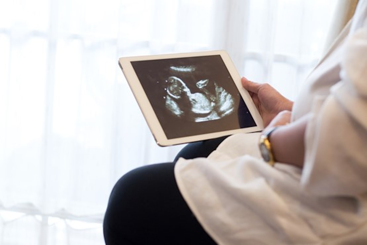
The benefits of 3D and 4D scans
Both 3D and 4D scans are considered just as safe as a 2D scan because the image is made up of sections of two-dimensional images converted into a picture.
However, the benefits of having more scans just for the fun of having another glimpse at your baby isn’t clear.
With 3D baby scansand 4D scans, you have the benefit of seeing your baby's skin covering her internal organs.
You may also get to see the shape of your baby’s mouth and nose, or see her yawn or stick her tongue out.
